Anatomy Muscles Pelvis : Muscles Of The Abdomen Lower Back And Pelvis / Cross the ls joint onto the trunk 2.. The muscles of the pelvic floor are collectively referred to as the levator ani and coccygeus muscles. Abdominal and pelvic anatomy encompasses the anatomy of all structures of the abdominal and pelvic cavities. The back of the abdomen consists of back muscles as well as the spine. Psoas consists of a pair of deep muscles (psoas major and iliacus) located on each side of the pelvis in the abdomen. See more ideas about anatomy, thoracic, basic image.
The greater pelvis is the space between the two wings of the ilium above the terminal line, while the lesser pelvis is the part of the pelvis below the terminal line. The largest of them is the most superficial muscle, the gluteus maximus. The levator ani muscles consist of three. This mri male pelvis axial cross sectional anatomy tool is absolutely free to use. Describe the muscles of pelvic diaphragm.
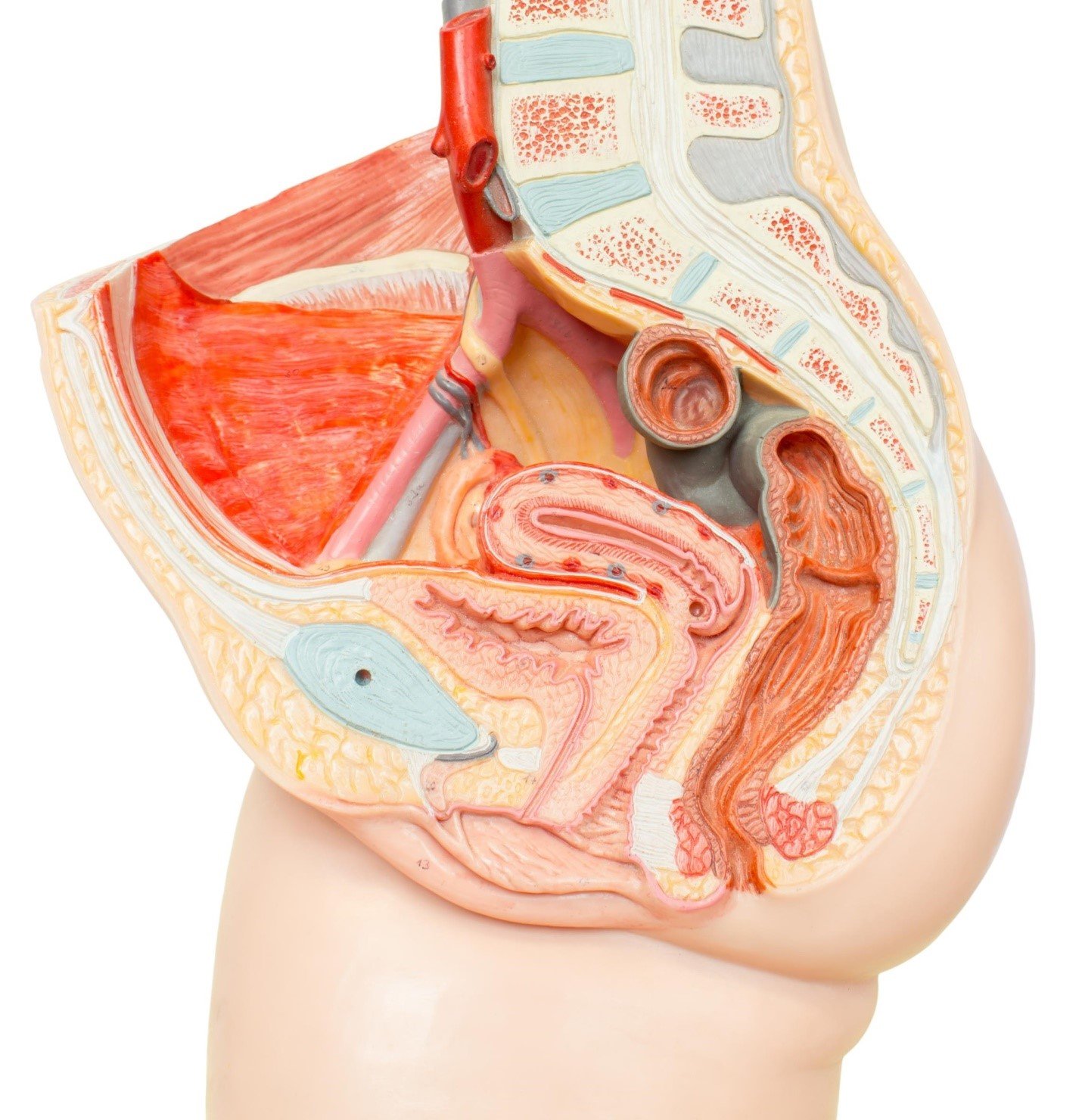
Female Pelvis With Detachable Pelvic Floor Muscles 12 Part Anatomy Models And Anatomical Charts from cdn3.volusion.com Included in this group are the adductor longus, adductor brevis, adductor magnus, pectineus, and gracilis muscles. The pelvic girdle is formed by a single hip bone. They have several functions, including helping to support the pelvic organs. It helps maintain erect posture, abducts the thigh, and rotates the thigh outward. Assoc prof craig hacking and dr tim luijkx et al. It originates from the pelvic outermost layer of the middle 3 sections of sacrum by 3 digitations. Cross the ls joint onto the trunk 2. The pubococcygeus (pc) muscle is the muscle that runs the show in pelvic floor health.
The muscles of the pelvic floor are collectively referred to as the levator ani and coccygeus muscles.
The pelvic girdle and pelvic spine. Below the gluteus maximus is the smaller gluteus medius. The adductor muscle group, also known as the groin muscles, is a group located on the medial side of the thigh. The iliacus muscle is part of a complex muscle system in the hip area that can function on its own or with other muscles. (1) the obturator internus and the piriformis, which are muscles of the lower extremity, and will be described with these (pages 476 and 477); Providing attachment for many muscles and ligaments used in locomotion; The muscles of the pelvis, hip and buttock anatomical chart shows how each muscle in this area of the body works with the others, and the various minor systems within the major ones. Psoas consists of a pair of deep muscles (psoas major and iliacus) located on each side of the pelvis in the abdomen. Continence, then pelvic muscle exercises may be effective. See more ideas about anatomy, thoracic, basic image. It is usually divided into two separate anatomic regions: It is composed of three separate paired muscles; The three muscles of the group—the iliacus, the psoas major, and the psoas minor—arise from different areas of your pelvis and lumbar spine to form a common attachment in your hip.
This mri male pelvis axial cross sectional anatomy tool is absolutely free to use. These muscles move the thigh toward the body's midline. To maintain the continence of urine and faeces. The muscles within the pelvis may be divided into two groups: The muscles of the pelvic floor are collectively referred to as the levator ani and coccygeus muscles.
Female Pelvic Floor 1 Anatomy And Pathophysiology Nursing Times from cdn.ps.emap.com The three muscles of the group—the iliacus, the psoas major, and the psoas minor—arise from different areas of your pelvis and lumbar spine to form a common attachment in your hip. See more ideas about anatomy, thoracic, basic image. The largest of them is the most superficial muscle, the gluteus maximus. (2) the levator ani and the coccygeus, which together form the pelvic diaphragm and are associated with the pelvic viscera. Attached to the pelvis are muscles of the buttocks, the lower back, and the thighs. The abdominal muscles lay in front of the fascia, and on the outermost layer is the skin. Assoc prof craig hacking and dr tim luijkx et al. On the other hand, if portions of those muscles are irretrievably lost, for example, due to complete.
It separates pelvis from the perineum.
Reviews the functional anatomy of the pelvic floor structures, the effects of age on urethral support and the urethral sphincter, and attempts to clarify Attached to the pelvis are muscles of the buttocks, the lower back, and the thighs. On the other hand, if portions of those muscles are irretrievably lost, for example, due to complete. Abdominal and pelvic anatomy encompasses the anatomy of all structures of the abdominal and pelvic cavities. These muscles move the thigh toward the body's midline. Muscles an important group of muscles in the pelvis is the pelvic floor. Included in this group are the adductor longus, adductor brevis, adductor magnus, pectineus, and gracilis muscles. Enclosing and protecting abdominopelvic and pelvic viscera. The pelvis is composed of the bony pelvis, pelvic floor, pelvic cavity, and the perineum. It helps maintain erect posture, abducts the thigh, and rotates the thigh outward. It originates from the pelvic outermost layer of the middle 3 sections of sacrum by 3 digitations. The abdominal muscles lay in front of the fascia, and on the outermost layer is the skin. The adductor muscle group, also known as the groin muscles, is a group located on the medial side of the thigh.
The pelvic girdle, also known as the hip bone, is composed of three fused bones: It is usually divided into two separate anatomic regions: These muscles move the thigh toward the body's midline. The iliopsoas muscle is a major hip flexor that also helps to move your spine. The classification of the two groups under a common heading is.
The Muscles That Control The Pelvic Floor Pericoach from www.pericoach.com To maintain the continence of urine and faeces. Cross the hip joint onto the thigh/leg 3. The pelvis is composed of the bony pelvis, pelvic floor, pelvic cavity, and the perineum. The pelvis is the lower portion of the trunk, located between the abdomen and the lower limbs. The muscles of the pelvis, hip and buttock anatomical chart shows how each muscle in this area of the body works with the others, and the various minor systems within the major ones. It's supplied by ventral rami of first and 2nd sacral nerves (s1, s2). Cross the ls joint onto the trunk 2. They have several functions, including helping to support the pelvic organs.
The pelvic girdle, also known as the hip bone, is composed of three fused bones:
Cross the ls joint onto the trunk 2. The muscles of the pelvis and hip control the vast range of movement of the legs and torso. As well as some basic images of disc pathology and stylised facet joint motion. Muscles that attach from the pelvis to the trunk and cross the lumbosacral joint muscles that attach from the pelvis to the thigh/leg and cross the hip joint pelvic floor muscles that are located wholly within the pelvis Arcus tendineus levator ani and the ischial spine It is composed of three separate paired muscles; The main function of the pelvic floor muscles are: Continence, then pelvic muscle exercises may be effective. The pelvic girdle, also known as the hip bone, is composed of three fused bones: These muscles move the thigh toward the body's midline. Large ligaments, tendons, and muscles around the hip joint hold the bones (ball and socket) in place and keep it from dislocating. See more ideas about anatomy, thoracic, basic image. (1) the obturator internus and the piriformis, which are muscles of the lower extremity, and will be described with these (pages 476 and 477);