Home » Without Label » Shoulder Anatomy Diagram - Shoulder Anatomy In Detail / Formerly called tendinitis, this is inflammation or irritation of a tendon that attaches to a bone.
Shoulder Anatomy Diagram - Shoulder Anatomy In Detail / Formerly called tendinitis, this is inflammation or irritation of a tendon that attaches to a bone.
Shoulder Anatomy Diagram - Shoulder Anatomy In Detail / Formerly called tendinitis, this is inflammation or irritation of a tendon that attaches to a bone.. Learn about these muscles, their origin and insertion points, and their functional anatomy. Four of them are found on the anterior aspect of the shoulder, whereas the rest are located on the shoulder's posterior aspect and in the back. The most common shoulder injuries involve the muscles, ligaments, cartilage, and tendons, rather than the bones. To learn more about how the shoulder muscles work, review the accompanying lesson called shoulder muscles: The shoulder is one of the most sophisticated and complicated joints of the human body.
Anatomy and injuries of the shoulder poster shoulder anatomy prints human body wall art picture body study canvas paintings exercise room gym decor 40x60cm no frame. This diagram depicts shoulder anatomy muscles diagram. The shoulder joint is the junction between the chest and the upper extremity. 17 photos of the diagram of shoulder muscles and tendons. Due to the inherent complexity of the shoulder joint, it is also particularly prone to problems.
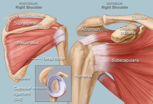
Anatomy Of The Shoulder Everything You Need To Know Dr Nabil Ebraheim Youtube from i.ytimg.com Muscles of the shoulder : Most people with rotator cuff injuries can recover with rest and physical therapy. The most flexible joint in the entire human body, our shoulder joint is formed by the union of the humerus, the scapula (or shoulder blade), and the clavicle (or collarbone). Find out in this anatomy of the shoulder quiz. For that reason, and because of the dexterity of the shoulder joint itself, the musculature of the shoulder is complex, ranging from massive prime mover muscles to finer stabilizer and fixator muscles. The muscles of the shoulder support and produce the movements of the shoulder girdle.they attach the appendicular skeleton of the upper limb to the axial skeleton of the trunk. Due to the inherent complexity of the shoulder joint, it is also particularly prone to problems. These muscles form the outer shape of the shoulder and underarm.
This diagram depicts shoulder instability xray photo.human anatomy diagrams show internal organs, cells, systems, conditions, symptoms and sickness information and/or tips for healthy living.
Ebraheim's educational animated video describes muscle anatomy of the shoulder girdle and anatomy of the shoulder joint.anatomy of the shoulder muscles a. There are many nerves and blood vessels that supply the muscles and bones of the shoulder. Plus, exercises for training them. For that reason, and because of the dexterity of the shoulder joint itself, the musculature of the shoulder is complex, ranging from massive prime mover muscles to finer stabilizer and fixator muscles. The shoulder, or glenohumeral joint, connects the upper arm to the chest. However, more serious injuries, such as complete rotator cuff tears, may require surgical repair. Formerly called tendinitis, this is inflammation or irritation of a tendon that attaches to a bone. Shoulder_anatomy_diagram_labrum 3/3 shoulder anatomy diagram labrum shoulder anatomy diagram labrum eventually, you will very discover a additional experience and talent by spending more cash. This diagram depicts shoulder instability xray photo.human anatomy diagrams show internal organs, cells, systems, conditions, symptoms and sickness information and/or tips for healthy living. The shoulder joint is formed where the humerus (upper arm bone) fits into the scapula (shoulder blade), like a ball and socket. To keep things simple, we can divide the shoulder into layers. Anatomy • free medical books. Human anatomy diagrams show internal organs, cells, systems, conditions, symptoms and sickness information and/or tips for healthy living.
Human anatomy diagrams show internal organs, cells, systems, conditions, symptoms and sickness information and/or tips for healthy living. Learn about these muscles, their origin and insertion points, and their functional anatomy. Numerous muscles help stabilize the three joints of. 3d tutorial on the anatomy of the shoulder joint from anatomyzone for more videos, 3d models and notes visit: Other important bones in the shoulder include:
Shoulder Human Anatomy Image Function Parts And More from img.webmd.com Discuss tha agaonist/antagonist relationship of muscles. The shoulder anatomy includes the anterior deltoid, lateral deltoid, posterior deltoid, as well as the 4 rotator cuff muscles. The shoulder joint (glenohumeral joint) is a ball and socket joint between the scapula and the humerus.it is the major joint connecting the upper limb to the trunk. To keep things simple, we can divide the shoulder into layers. These muscles form the outer shape of the shoulder and underarm. The shoulder, or glenohumeral joint, connects the upper arm to the chest. Starting with what is deepest, it goes: The shoulder joint is formed where the humerus (upper arm bone) fits into the scapula (shoulder blade), like a ball and socket.
Neck and shoulder anatomy diagram :
It is one of the most mobile joints in the human body, at the cost of joint stability. Two joints are at the shoulder. Ebraheim's educational animated video describes muscle anatomy of the shoulder girdle and anatomy of the shoulder joint.anatomy of the shoulder muscles a. These muscles form the outer shape of the shoulder and underarm. Learn their origins/insertions, functions & exercises. To learn more about how the shoulder muscles work, review the accompanying lesson called shoulder muscles: Anatomy and injuries of the shoulder anatomical chart. The muscles of the shoulder support and produce the movements of the shoulder girdle.they attach the appendicular skeleton of the upper limb to the axial skeleton of the trunk. However, more serious injuries, such as complete rotator cuff tears, may require surgical repair. Find out in this anatomy of the shoulder quiz. Male shoulder ligaments and biceps muscles isolated in skeleton labeled chart on white labeled human anatomy diagram of male shoulder ligaments, connective tissue and biceps muscles isolated within the skeletal system frontal anterior view on a white background. The following is an overview of the shoulder muscle anatomy. The shoulder has about eight muscles that attach to the scapula, humerus, and clavicle.
There are many nerves and blood vessels that supply the muscles and bones of the shoulder. Name this muscle that elevates the shoulder. Bones in shoulder, ligaments of the shoulder joint, parts of the shoulder joint, shoulder anatomy, shoulder joints and muscles, shoulder structure anatomy, shoulder tendon anatomy, shoulder tendons ligaments, human muscles, bones in shoulder, ligaments of the shoulder joint, parts of. This diagram depicts shoulder anatomy muscles diagram. Browse 3,854 shoulder anatomy stock photos and images available, or search for shoulder joint or rotator cuff to find more great stock photos and pictures.
Shoulder Anatomy Image Anatomy Drawing Diagram from medicalartlibrary.com The shoulder joint (glenohumeral joint) is a ball and socket joint between the scapula and the humerus.it is the major joint connecting the upper limb to the trunk. Ebraheim's educational animated video describes muscle anatomy of the shoulder girdle and anatomy of the shoulder joint.anatomy of the shoulder muscles a. Shoulder_anatomy_diagram_labrum 3/3 shoulder anatomy diagram labrum shoulder anatomy diagram labrum eventually, you will very discover a additional experience and talent by spending more cash. Muscles of the shoulder : This lesson covers the following objectives, along with. Plus, exercises for training them. The most common shoulder injuries involve the muscles, ligaments, cartilage, and tendons, rather than the bones. Last update september 3, 2020.
Anatomy and injuries of the shoulder poster shoulder anatomy prints human body wall art picture body study canvas paintings exercise room gym decor 40x60cm no frame.
The shoulder joint is the junction between the chest and the upper extremity. Learn about these muscles, their origin and insertion points, and their functional anatomy. The upper arm bone, called the humerus, is connected to the body via the shoulder blade, which possesses the latin name scapula. Anatomy • free medical books. The following is an overview of the shoulder muscle anatomy. Human anatomy diagrams show internal organs, cells, systems, conditions, symptoms and sickness information and/or tips for healthy living. Name this muscle the largest of the shoulder group. Numerous muscles help stabilize the three joints of. However, more serious injuries, such as complete rotator cuff tears, may require surgical repair. Anatomy and injuries of the shoulder poster shoulder anatomy prints human body wall art picture body study canvas paintings exercise room gym decor 40x60cm no frame. Male shoulder ligaments and biceps muscles isolated in skeleton labeled chart on white labeled human anatomy diagram of male shoulder ligaments, connective tissue and biceps muscles isolated within the skeletal system frontal anterior view on a white background. Ebraheim's educational animated video describes muscle anatomy of the shoulder girdle and anatomy of the shoulder joint.anatomy of the shoulder muscles a. It causes pain in the area just outside the joint.